What Is Squamous Cell Carcinoma You Ask?
History: "Mondo" is a 7 year old Clydesdale gelding who was rescued by his current owners approximately a year and a half ago. Patient was malnourished and obviously neglected. He also had a yellowish discharge from his right eye.

August 2010: At the time
of the rescue, a routine physical examination was performed by Dr. Callie Cuthbertson. The right eye was draining
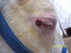
a yellowish discharge and upon closer inspection, a small mass was noted on the
third eyelid and another more obvious mass was seen on the lateral limbus
(where the white of the eye meets the cornea in the outside corner of the eye). The mass was no more than 2-3mm in
diameter. At that time, a diagnosis of
squamous cell carcinoma (SCC) of the right eye was made. This conclusion was reached because of the
gross physical appearance of the mass in conjunction with the fact that SCC is
the most common tumor of the equine eye/eyelid. While there are other types of cancer that affect the equine eye, including sarcoids, SCC is the most frequently seen. It was recommended at that time to pursue a treatment plan, due to the
fact that ocular SCC tends to be very fast growing and locally invasive.
No photograph of Mondo's initial presentation was available. This is a photo of a very similarly presenting SCC.
No photograph of Mondo's initial presentation was available. This is a photo of a very similarly presenting SCC.

January 2011: Dr. Callie was called out to recheck the eye about 6 months later. The mass at the limbus had grown significantly, quadrupling in size. Luckily, this SCC was relatively slow growing; many cases of ophthalmic SCC grow much more quickly. At that time, it was decided that the patient would be seen by a local veterinary ophthalmologist.

February 2011: Mondo was placed under general anesthesia in the field. The tumor was debulked (as much as possible was removed) and cisplatnin beads (small beads impregnated with chemotherapy) were implanted in the surrounding tissue by Dr. Blair, the ophthalmologist. The third eyelid was removed as well as a precaution.
March 2011: Mondo was sedated and treated with cryotherapy by Dr. Blair about 1 month after the surgical debulking. This entails freezing off any unwanted tissue, specifically the tumor.
March 2011: Mondo was sedated and treated with cryotherapy by Dr. Blair about 1 month after the surgical debulking. This entails freezing off any unwanted tissue, specifically the tumor.

September 2011: Patient was rechecked by Dr. Callie. Tumor had not only grown back, but increased significantly in size. No normal cornea was visible, as the tumor had grown to cover the surface of the eye. At this time, it was decided that the patient’s best chance for survival was to enucleate (remove) the eye.

October 2011: Patient was again anesthetized in the field. This time the right eye and the surrounding tissues/structures were entirely removed. The patient recovered extremely well from the surgery.
The take-home message from this case is that ophthalmic cancers must be caught very early and treated extremely aggressively. There are many different treatment options available when dealing with ophthalmic tumors, including surgical excision, local chemotherapy, cryotherapy and enucleation. An individualized treatment plan will be tailored for each horse. In this case, Mondo no longer has his eye, but he does have his life, which is a very happy and healthy one.
For additional information and details about SCC and other tumors of the equine eye, or if you have any concerns regarding your horse, please contact us.
Additional Ophthalmic SCC Cases
Below are some photos of less lucky horses suffering from SCC of the eye/surrounding structures. These patients were treated with various appropriate therapeutic options. These pictures are included to demonstrate the seriousness of this disease. Please be aware that these photos are graphic.

This is the initial presentation for a patient diagnosed with SCC of the lower eyelid. This tumor was actually inside and involving the internal structures of the lid.

This is the same patient 6 months later. This patient had been treated with a locally injected chemotherapeutic agent. This patient was euthanized.
This is the initial presentation of a SCC along the lower lid margin. No internal structures were involved. This small tumor was surgically excised from the lower lid and then treated with cryotherapy.

This is about 16 months after cryotherapy treatment. Note the extensive infiltration of tumor into the surrounding structures of the head. The right side of the head is extremely distorted when compared with the patient's left. This patient was also euthanized.
Owner permission was given prior to this case being posted online. The patients' name were changed to comply with confidentiality laws and to protect the innocent.
Dover Equine Veterinary
P.O. Box 246 | Aylett, VA 23009
804.333.0333 | 804.769.2433
doverequinevet@gmail.com