December's Case
History: "Mr. Pickle" is a 3 year old Morgan gelding. He resides in a large field with a large open barn for shelter and two other horses. One Sunday, the owner was picking the patient's hooves prior to riding. She noticed what appeared to be a metallic object sticking out of his right hind frog (it should be noted that the patient was only slightly lame - this might not have been found immediately if not for her vigilance). She left it in place, which was an excellent decision, and paged the emergency line.
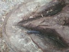
Physical Exam: Mr. Pickle's vital signs were all within normal limits. The only abnormalities noted were a grade III/V lameness on the right hind limb and 2.5cm of metal implement protruding from the lateral sulcus of the frog, approximately 3cm behind the point of the frog.
Radiographs: It was very important to find out what musculoskeletal structures, if any, are damaged by the foreign body. Below are just a few of the radiographs, or xrays, taken that day. Luckily, the drill bit is directed up and back at a low angle, resulting in it missing the vital structures listed below.
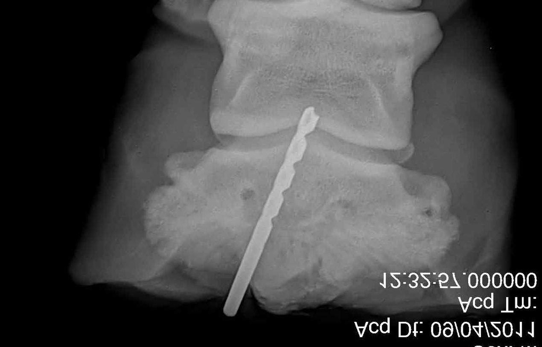
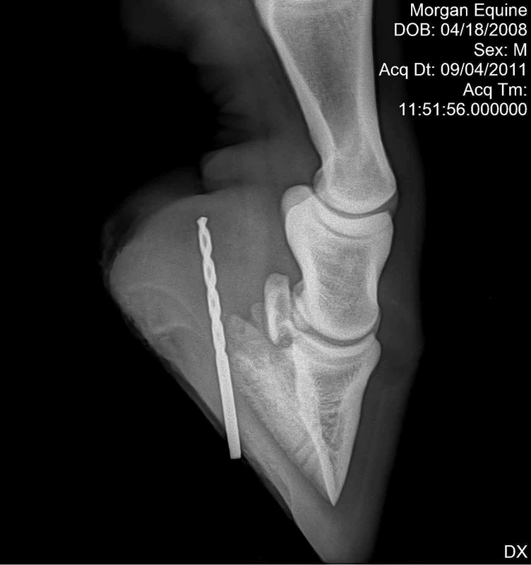
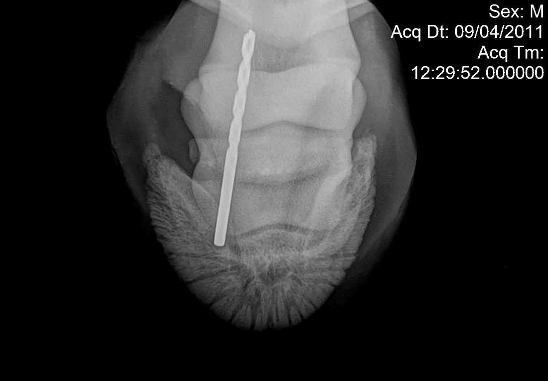
Differential Diagnosis: The actual diagnosis in this case is relatively straightforward - obviously there is a puncture wound due to a penetrating foreign body. The most important part of the diagnosis is where that foreign body went. The potentially affected structures include the navicular bone, P3, the joints, the navicular bursa and/or the deep digital flexor tendon. In this case, Mr. Pickle was extremely lucky, and the bottom of P3 was the only structure that may have been involved by being scratched but not penetrated by the drill bit.
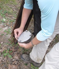
Treatment: Now that we know exactly where the foreign body went and what structures were, or weren't, affected, it is time to remove the foreign body and develop a treatment plan. The most important treatment strategies include maintaining the cleanliness of the wound, controlling inflammation and pain, and combating infection. The location of the wound does not lend itself to remaining clean and will be very prone to being packed full of dirt and therefore bacteria. The wound was cleaned daily and a hospital plate was applied compliments of the farrier, Joe Cuthbertson of Cuthbertson Farrier Service. The hospital plate (pictured left) allows for easy access to the bottom of the foot for cleaning while being simple to remove from the foot but making an easy seal over the sole. The patient was placed on an NSAID to ease pain and inflammation. In order to control any potential infection, a two-pronged approach was taken. Firstly, he was placed on an oral antibiotic. Secondly, a procedure called "regional limb perfusion" was performed daily. With this, a tourniquet is placed on the affected limb and below it a catheter is inserted into a vein and used to administer an antibiotic. The idea is that a concentrated level of antibiotic is administered directly to the area that most needs it.
Prognosis: Mr. Pickle's prognosis is very good to excellent for return to normal function. This is largely due to the fact that the injury was discovered so early and treatment was immediately sought from the veterinarian.
Owner permission was given prior to this case being posted online. The patient's name was changed to comply with confidentiality laws and to protect the innocent.
Dover Equine Veterinary
P.O. Box 246 | Aylett, VA 23009
804.333.0333 | 804.769.2433